

News
Accelerating Cancer Research: How A Top Tech Microscope Can Fast-Track Drug Design
Cryo-electron microscopy (cryo-EM) has become a powerful tool in both fundamental biology as well as drug discovery and development. To accelerate cancer research, the Oncode Accelerator program recently invested in a state-of-the-art Glacios cryo-EM microscope. Dr. Steffen Brünle, Assistant Professor at Leiden University, explains how this technology is helping scientists to fast-track their cancer research.
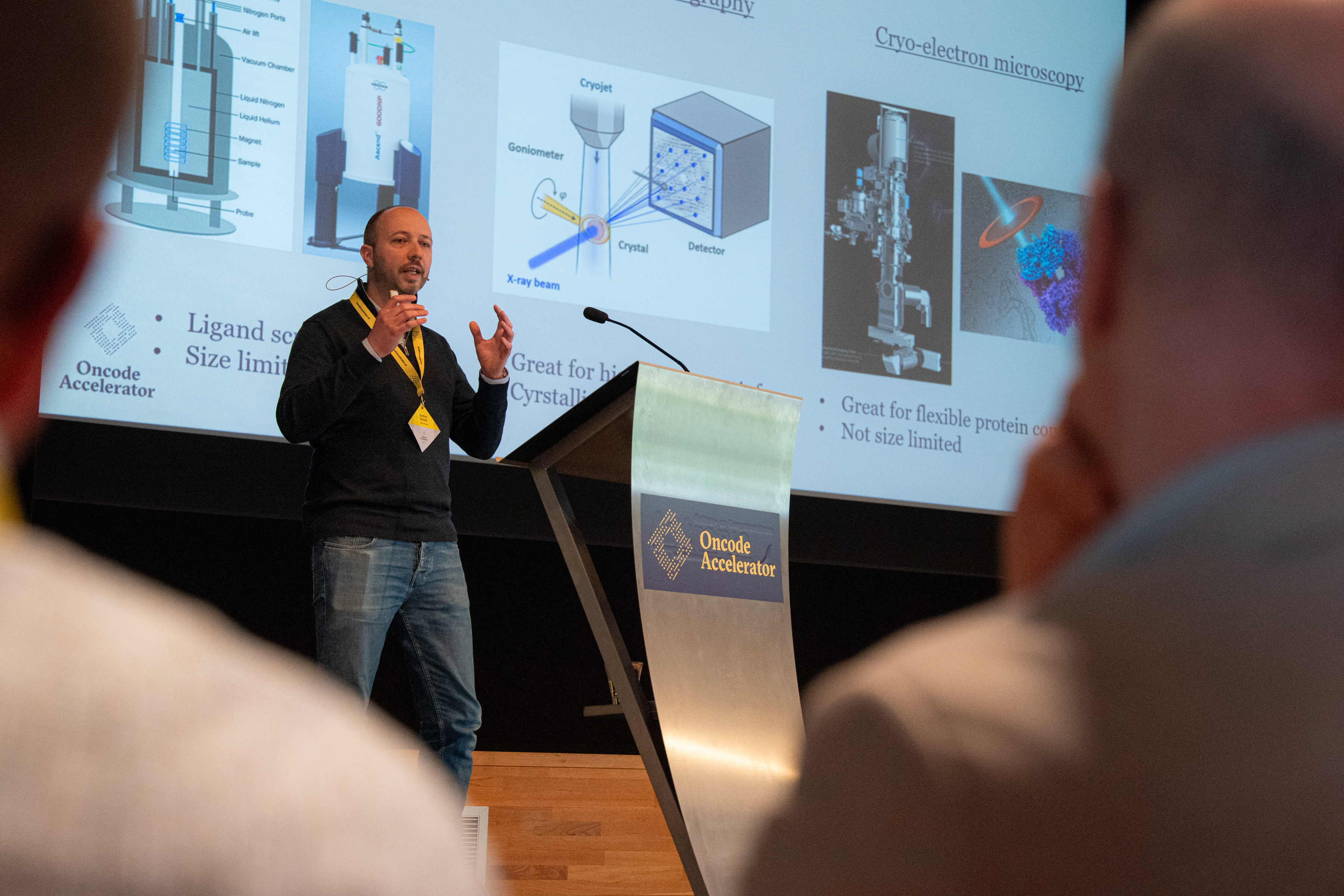
Structure matters, in daily life and in biology. For instance, think of a water bottle. You understand how to use it just by looking at it—where to pour water, how to close it, and how it holds liquid. This simple idea also applies to biology.
“If you understand the structure of a molecule, you can start to understand the function,” explains Steffen Brünle, Assistant Professor at Leiden University and member of the Oncode Accelerator Small Molecules Workstream. “So, if you know how a protein looks in 3D, you can begin to understand how it works. It’s like knowing if a water bottle has a screw cap or a pop-top—once you know how it looks, you can understand how to interact with it.”
Cryo-EM is one of several techniques used to study molecular structures. It has revolutionized the field of structural biology by making it easier for scientists to get clear, high-resolution images of tiny biological molecules much faster than before. The importance of this method was recognized with the Nobel Prize in Chemistry in 2017. The winning scientists developed a way to freeze molecules in place so they could be imaged in striking detail—down to the level of individual atoms.
Where Structure Meets Strategy in Drug Discovery and Development
Steffen Brünle became fascinated with structural biology early in his career, and it brought him from Germany to Switzerland, and eventually to the Netherlands. At first, he specialized mainly in x-ray crystallography (the technique that was used to define DNA’s double-helix structure), but he later also added cryo-EM to his toolkit.
“I aim to use both methods as a combinatory approach to achieve the same goal: to better understand protein interactions, and how we could design drugs to influence those interactions,” Brünle explains.
Brünle’s research group at Leiden University studies G protein-coupled receptors (GPCRs), a family of proteins that plays a crucial role in how cells respond to different signals. GPCRs are also important targets in drug discovery – many drugs work by targeting these receptors. Using cryo-EM alongside x-ray crystallography, his group is uncovering how different molecules interact with GPCRs in different ways, to trigger different outcomes.
“Depending on where a molecule binds to the receptor, it can cause completely different responses in the cell,” says Brünle. “If we can understand that process better, we can design drugs that are more precise—and thereby limit side effects for patients.”
Using Top Tech to Fast-Track Research
The Glacios microscope recently purchased through Oncode Accelerator can support scientists in achieving this goal of designing precision medicines faster. Brünle explains:
Steffen Brünle, Assistant Professor at Leiden University and member of the Oncode Accelerator Small Molecules WorkstreamThis microscope is very well developed. It has the latest detector technology and allows us to very rapidly gain structural insights into our samples at a fraction of the cost compared to other microscopes, and in only a few hours.
In the past, cryo-EM scientists used a time-consuming, three-step process to assess and optimize sample quality and gain detailed information about the molecules under study. This process also involved three different microscopes. First, a relatively inexpensive and low-powered microscope was used for a rough assessment of the biological sample. Then, a more expensive microscope (known as a 200kV microscope) would be used to optimize the sample. Only then would a very expensive, high-powered microscope (what scientists call a 300kV microscope) finally be used to take the images needed to create a picture of the molecule of interest at atomic detail.
Now, the Glacios – a 200kV microscope that makes use of the latest innovations in electron microscopy – has simplified that long process into a single, streamlined step.
“Now,” explains Brünle, “Sample assessment using the inexpensive and low-powered microscope is obsolete, and going to the most expensive type of microscope, a 300kV microscope, is optional. The Glacios is a highly capable 200kV instrument that already allows us to perform high-resolution structure determination, allowing us to accelerate our science considerably.”
The Glacios microscope's efficiency and cost-effectiveness make it a valuable investment within the Oncode Accelerator ecosystem – in line with our mission to collaboratively optimize and accelerate the development of new cancer therapies for the benefit of patients of all ages.
The Power of Collaborative Science
Alongside top tech, the right kind of collaboration can also accelerate drug discovery. Embedding cryo-EM expertise and technologies into Oncode Accelerator’s research ecosystem can boost the ability of scientists to rapidly design and optimize small molecule cancer therapeutics. Brünle explains:
Steffen Brünle, Assistant Professor at Leiden University and member of the Oncode Accelerator Small Molecules WorkstreamBeing based in Leiden and within the Oncode Accelerator program is ideal for my research. This ecosystem brings together experts in medicine, chemistry, and other drug discovery-related disciplines, which creates a great research environment.
This kind of collaboration, which blends complementary expertise, can help scientists to progress much faster. For instance, teams working on developing cancer drugs can use cryo-EM to see exactly how different compounds could interact with target proteins – and then once they have that structural information, efficiently fine-tune those compounds to make them work better.
“There are a lot of collaborative projects going on,” says Brünle. “Many people are now developing small molecules, for instance as potential cancer therapies, and there is a lot of interest in using cryo-EM to see how those molecules interact with a protein of interest. Once you have that information, then you can use a structure-based drug discovery approach to optimize your drug candidate.”
Steffen Brünle's work underlines how combining cutting-edge technology with strong collaboration can accelerate scientific discovery and development. By using top tech like the Glacios cryo-EM microscope, researchers can accelerate towards developing more effective, targeted treatments that could eventually benefit cancer patients.
Leiden University was founded in 1575 and is one of the leading international research universities in Europe. The University has seven faculties and a campus in both Leiden and The Hague. Our motto is Praesidium Libertatis – Bastion of Freedom.
Through our research, we work towards a fundamental understanding of the world and people around us. We use that knowledge to make the world a safe, healthy, sustainable, prosperous and just place.
Society is facing complex issues, such as the climate crisis, wars, food shortages and increasing numbers of displaced people. As a broad-based university, it is our strength to help find solutions to such issues. Because to get to grips with complex problems we need insights from different perspectives and to transcend disciplines. Science and innovation cannot exist without each other, and our research also inspires our teaching. Through these cross-pollinations, we shape our mission from society.
Leiden University is an international, broad-based university. Our academics conduct research in five broad clusters: